microscopist
⏳ Unreviewed profession0
daily views
0 total
Microscopy is the technical field of using microscopes to view subjects too small to be seen with the naked eye (objects that are not within the resolution range of the normal eye). There are three well-known branches of microscopy: optical, electron, and scanning probe microscopy, along with the emerging field of X-ray microscopy.
Optical microscopy and electron microscopy involve the diffraction, reflection, or refraction of electromagnetic radiation/electron beams interacting with the specimen, and the collection of the scattered radiation or another signal in order to create an image. This process may be carried out by wide-field irradiation of the sample (for example standard light microscopy and transmission electron microscopy) or by scanning a fine beam over the sample (for example...
Current Images

Main article image
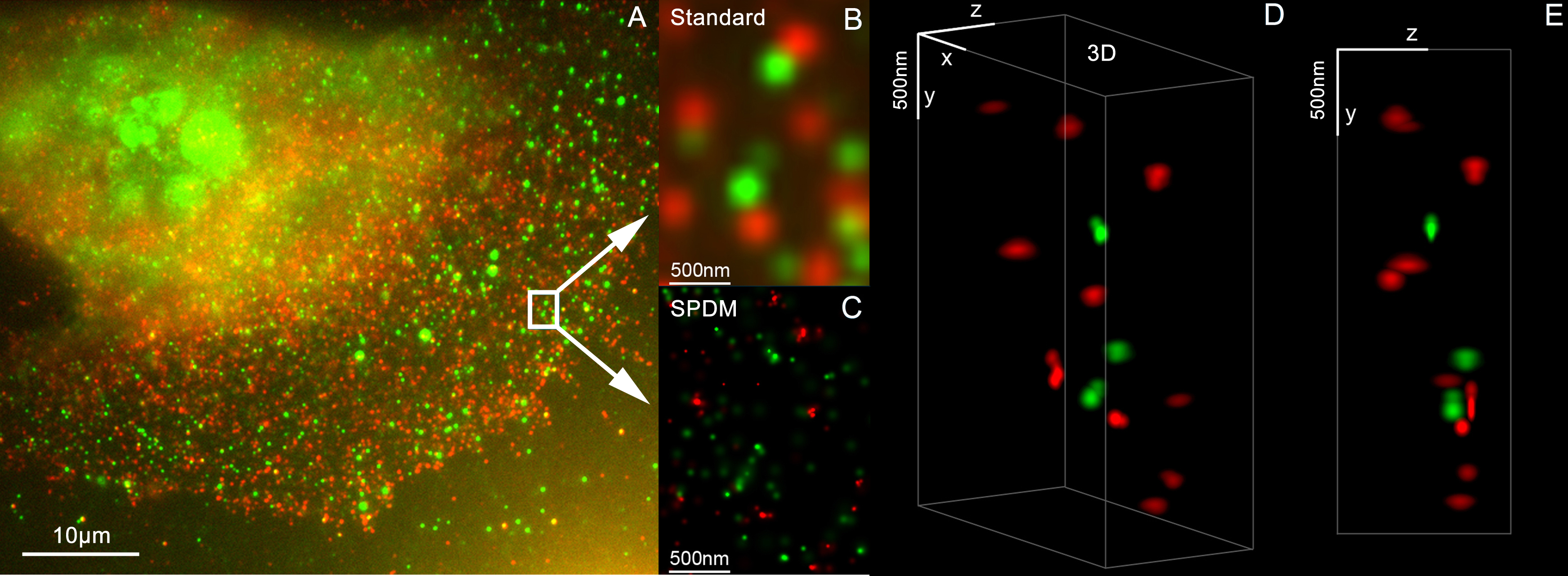
3D Dual Color Super Resolution Microscopy Cremer 2010.png
3D Dual Color Super Resolution Microscopy by combining localization microscopy SPDM with spatially m...
3D Dual Color Super Resolution Microscopy by combining localization microscopy SPDM with spatially m...

Anther of thale cress (Arabidopsis thaliana), an artefact.jpg
Confocal laser scanning fluorescence micrograph of thale cress anther (part of stamen). The picture ...
Confocal laser scanning fluorescence micrograph of thale cress anther (part of stamen). The picture ...

Anthonie van Leeuwenhoek (1632-1723). Natuurkundige te Delft Rijksmuseum SK-A-957.jpeg
Portrait of Anthonie van Leeuwenhoek (1632-1723), natural philosopher, in Delft. Knee piece, sittin...
Portrait of Anthonie van Leeuwenhoek (1632-1723), natural philosopher, in Delft. Knee piece, sittin...

FernProthallium400x.jpg
Microscopic view of Fern Prothallium
Microscopic view of Fern Prothallium

Housebeemouth100x.jpg
House Bee Mouth Magnified 100 times
House Bee Mouth Magnified 100 times

HumanRBCsPAM.png
Human red blood cells on a glass slide fixed with methanol.
Human red blood cells on a glass slide fixed with methanol.
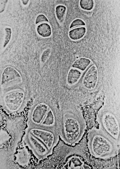
Hypertrophic Zone of Epiphyseal Plate.jpg
Light micrograph of an undecalcified epiphyseal plate that is displaying the hypertrophic zone with ...
Light micrograph of an undecalcified epiphyseal plate that is displaying the hypertrophic zone with ...
Loupe-binoculaire-p1030891.jpg
binocular microscope
binocular microscope

Microscopic observation, Микроскопирање.jpg
Microscopic observation
Microscopic observation
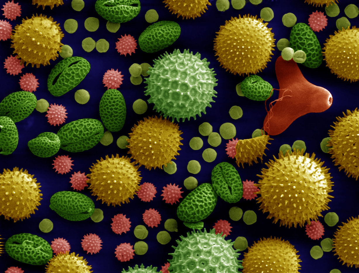
Misc pollen colorized.jpg
Pollen from a variety of common plants: sunflower (Helianthus annuus, small spiky sphericals, colori...
Pollen from a variety of common plants: sunflower (Helianthus annuus, small spiky sphericals, colori...
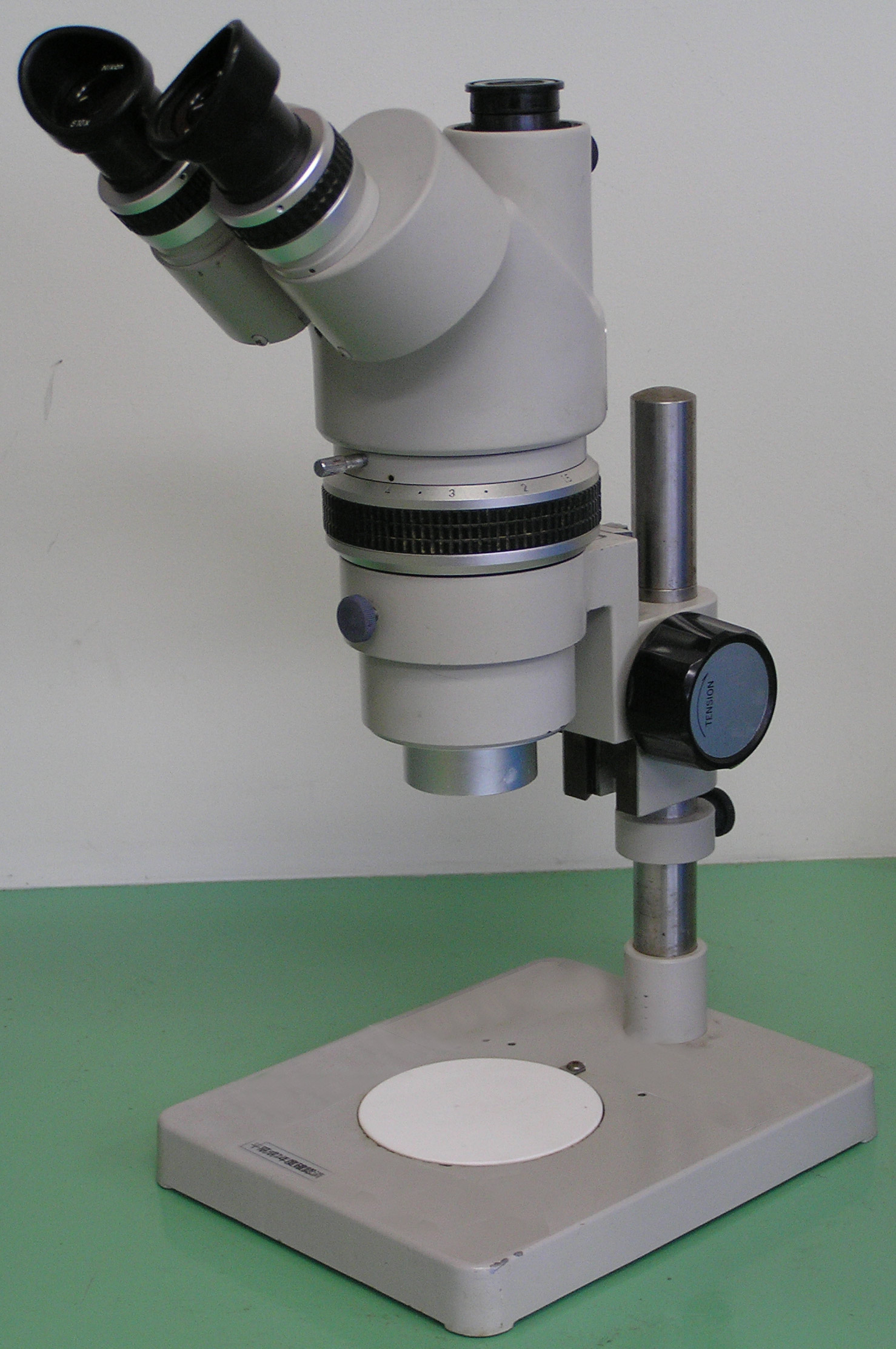
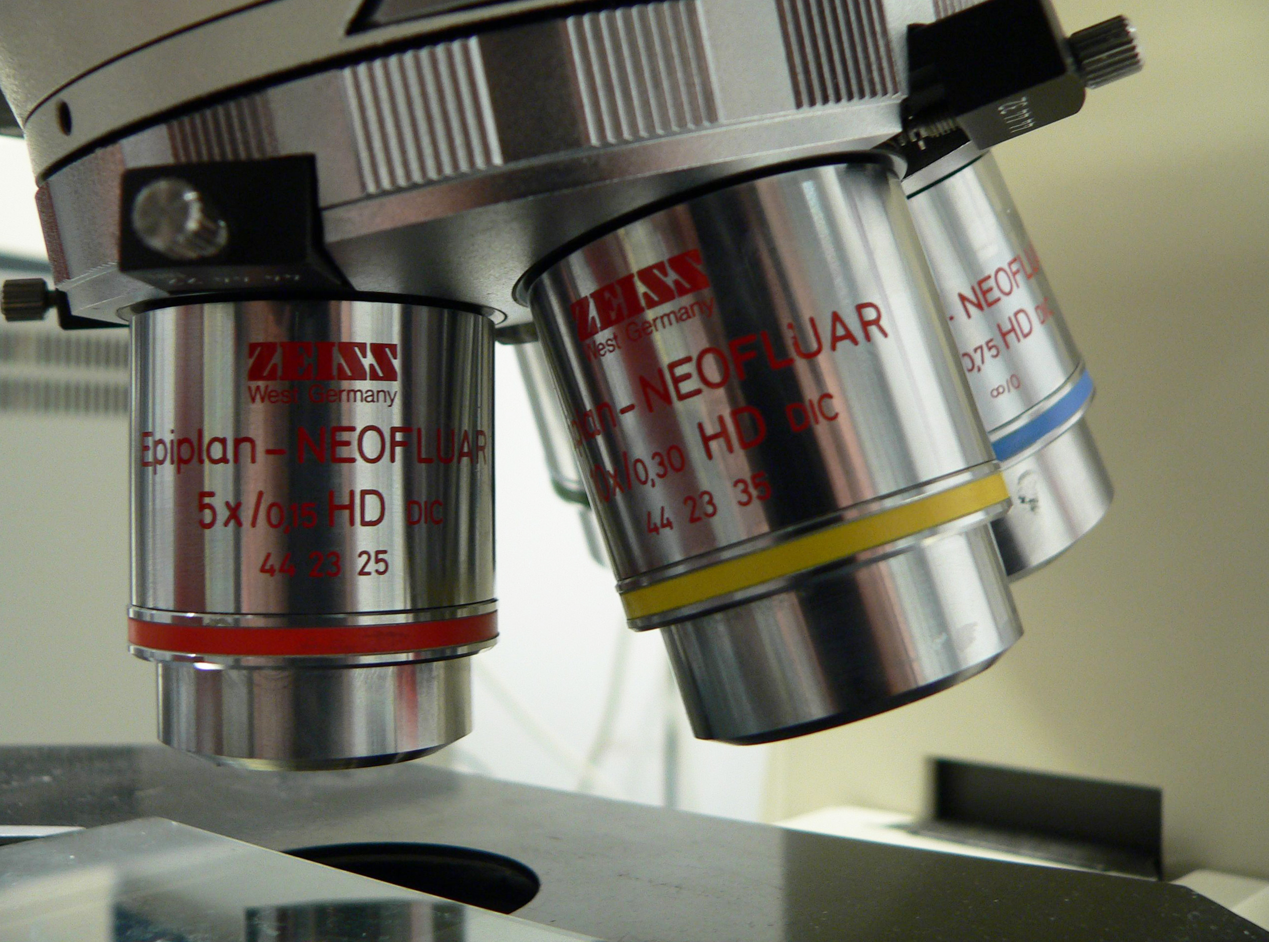
Optical stereo microscope nikon smz10.jpg
typical optical stereo microscope for academic use in 1980-1990s,Nikon SMZ-10
typical optical stereo microscope for academic use in 1980-1990s,Nikon SMZ-10

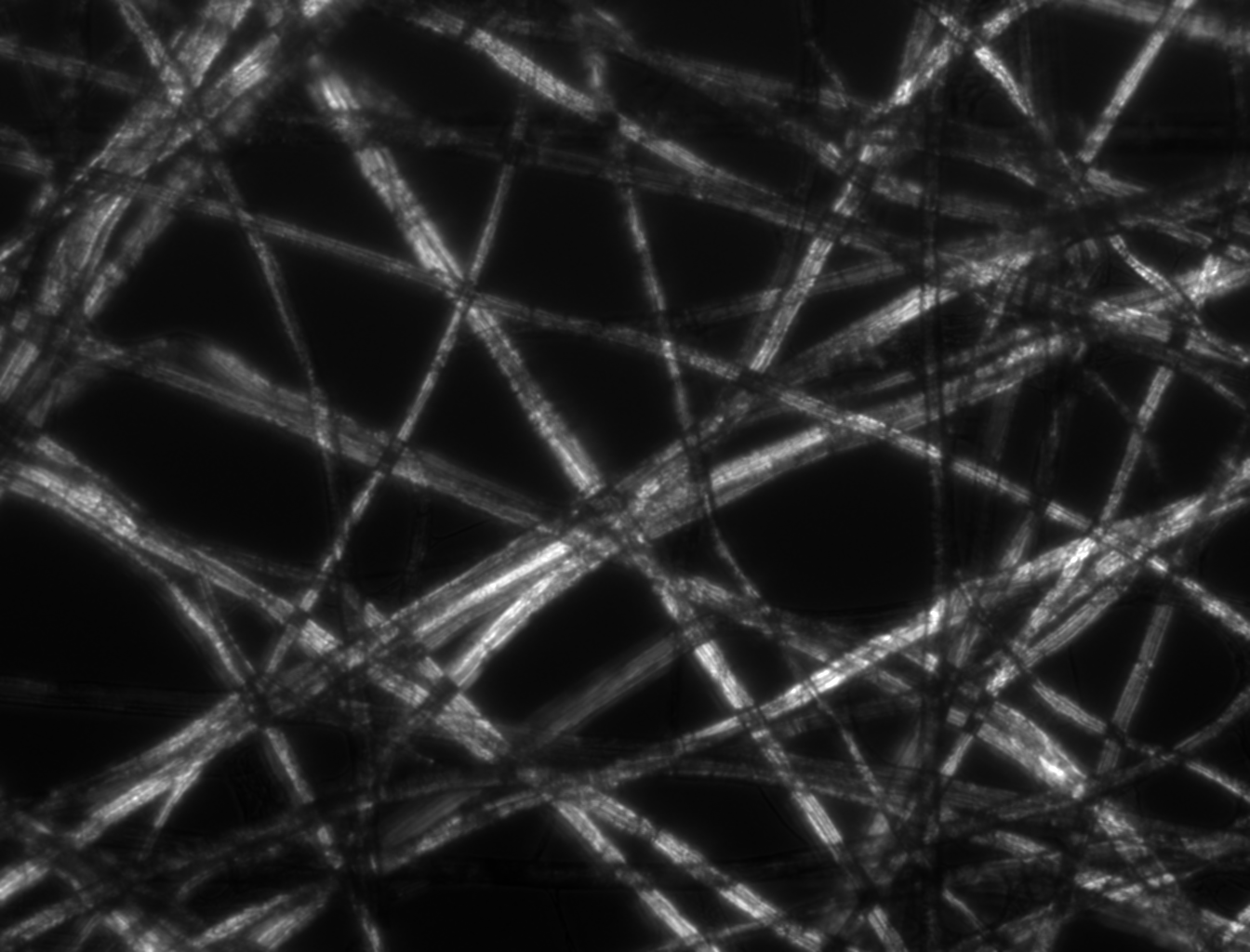
Paper Micrograph Bright.png
Micrograph of Whatman lens tissue paper. Bright field illumination. 10x magnification, 1.559 μm/px.
Micrograph of Whatman lens tissue paper. Bright field illumination. 10x magnification, 1.559 μm/px.
Paper Micrograph Cross-Polarised.png
Micrograph of Whatman lens tissue paper. Cross-polarised illumination. 10x magnification, 1.559 μm/p...
Micrograph of Whatman lens tissue paper. Cross-polarised illumination. 10x magnification, 1.559 μm/p...

Paper Micrograph Dark.png
Micrograph of Whatman lens tissue paper. Dark field illumination. 10x magnification, 1.559 μm/px.
Micrograph of Whatman lens tissue paper. Dark field illumination. 10x magnification, 1.559 μm/px.

Paper Micrograph Phase.png
Micrograph of Whatman lens tissue paper. Phase contrast illumination. 10x magnification, 1.559 μm/px...
Micrograph of Whatman lens tissue paper. Phase contrast illumination. 10x magnification, 1.559 μm/px...
People icon.svg
People icon
People icon

Phase-Phase Contrast.jpg
Comparison of digital holographic microscopy (DHM) phase shift (left) and a phase contrast microscop...
Comparison of digital holographic microscopy (DHM) phase shift (left) and a phase contrast microscop...

Rabbitttestis100x2.jpg
Microscopic view of rabbit testis, magnified 100 times.
Microscopic view of rabbit testis, magnified 100 times.

Rheinberg 6.jpg
Microscopic Photography of a Diatom with Rheinberg Illumination.
Microscopic Photography of a Diatom with Rheinberg Illumination.

RiceStemcs400x1.jpg
Rice stem mangified 400 times
Rice stem mangified 400 times
Search Openverse for Replacements
Presets: